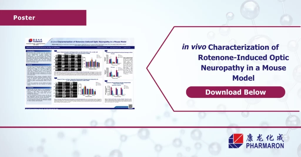
in vivo Characterization of Rotenone-Induced Optic Neuropathy in a Mouse Model
This study evaluates a mouse model of Leber’s Hereditary Optic Neuropathy (LHON) using intravitreal Rotenone to induce dose- and time-dependent optic nerve damage. While the model’s molecular mechanisms are known, its in vivo features had not been fully defined.
Key highlights from our optic neuropathy poster:
- Retinal changes: SD-OCT showed early GCC thickening (Day 4) followed by thinning (Day 14), indicating progression from inflammation to neurodegeneration.
- Functional loss: High-dose Rotenone reduced PERG amplitudes at Day 4, suggesting early retinal dysfunction.
- Therapeutic relevance: This model provides a rapid and quantifiable way to screen potential treatments for optic neuropathies like LHON.
Our results confirm that SD-OCT and PERG are effective tools for monitoring retinal structure and function in vivo.